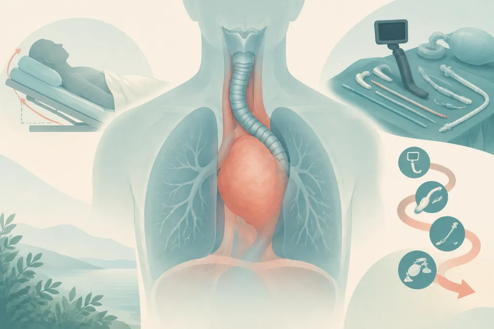
Imagine a patient who is comfortable sitting forward but coughs and becomes breathless when asked to lie flat. Computed tomography (CT) shows a narrowed trachea and a small pericardial effusion. The anesthetic risk is present before anyone gives a drug.
Anesthesia for an anterior mediastinal mass should be planned around the structures compressed, the patient’s symptoms and the rescue options that can actually be delivered. Preserving spontaneous ventilation is often prudent, particularly in children with significant compression, but it is not a universal rule or a rescue plan by itself.
Why an awake patient can still be precarious
An awake patient may compensate through posture, respiratory effort and muscle tone. Anesthesia can reduce functional residual capacity and airway tone; lying supine may change the mass’s relationship to the airway, heart and great vessels. A tracheal tube may secure the upper airway yet do nothing for a compressed carina or main bronchus below its tip.
Cardiovascular compromise has a different mechanism. Compression of the superior vena cava can reduce venous return. Compression of the right ventricle or pulmonary artery can obstruct right-sided outflow, while compression of cardiac inflow can impair filling. Positive-pressure ventilation may further reduce preload, particularly when venous return is already compromised or tamponade physiology is present. Airway and cardiovascular collapse can therefore occur separately or together. A normal oxygen saturation while awake does not rule either out.
Build the risk picture before choosing the anesthetic
Ask what happens in different positions. Orthopnea, cough or stridor when supine, wheeze, cyanosis, syncope or presyncope, positional hypotension, and swelling of the face, neck or arms all matter. Ask which position brings relief; it may be sitting, lateral or prone. Do not reproduce symptoms simply to complete an examination.
Review recent CT images, not only the report. Map the narrowest airway segment and its length, identify carinal or main-bronchial compression, and look for displacement or compression of major vessels and the heart. Transthoracic echocardiography helps assess pericardial effusion, tamponade physiology, ventricular function and compression of cardiac inflow or outflow. In adults, pulmonary-function findings may add information about postoperative respiratory risk, but they do not replace anatomical imaging.
A threshold is a warning, not a verdict. In an adult cohort, tracheal compression greater than 50% was associated with postoperative respiratory complications; the study did not make that figure a standalone prediction of intraoperative collapse. Pediatric literature uses different thresholds and may measure cross-sectional area rather than diameter. Those measurements are not interchangeable. Treat the degree and location of compression as part of a pattern that includes symptoms, bronchial involvement and cardiovascular findings.
An asymptomatic patient can still have important compression, especially a child who may not describe positional symptoms reliably. Conversely, a striking CT image does not by itself dictate one anesthetic technique. The strong candidate integrates the history, anatomy and physiology rather than reciting a cutoff.
First ask whether general anesthesia is needed
A diagnostic biopsy is not automatically a general anesthetic. Look for an accessible peripheral lymph node, pleural fluid or another site that can provide tissue under local anesthesia. Depending on the patient and diagnostic question, image-guided biopsy, bone-marrow sampling or another limited procedure may also avoid deep anesthesia. Sedation is not risk-free: it can reduce airway tone and ventilatory drive, so the location, monitoring and rescue resources still matter.
Discuss the procedure with anesthesia, oncology, radiology and the relevant surgical or airway teams. For a patient with substantial risk, the operating room may be safer than a remote location because experienced staff, airway equipment and emergency support are more readily available. If general anesthesia is unavoidable, agree the patient’s best position, airway approach and escalation plan in advance.
Induction is a staged choice, not a slogan
For a patient with severe positional symptoms, major airway compression or important pediatric risk, maintaining spontaneous ventilation and avoiding deep anesthesia until the team is ready to proceed is often a prudent starting point. Use the position the patient tolerates. If a tracheal tube is planned, check whether it can pass beyond the compressed segment. Awake intubation may protect the upper airway, but it cannot bypass a distal obstruction the tube cannot reach.
Newer adult evidence qualifies the traditional warning about positive pressure and paralysis. In a prospective observational study, 17 adults with large mediastinal masses underwent awake intubation followed by staged induction and bronchoscopic airway assessment. Airway patency scores did not worsen with positive-pressure ventilation and improved, on average, after neuromuscular blockade; no patient experienced airway collapse or difficult ventilation during the study. This evidence argues against treating paralysis as an absolute prohibition in every adult. It does not establish that an ordinary, unstaged induction is safe, settle cardiovascular risk or define an approach for children.
Suppose a child has orthopnea, main-bronchial compression and a pericardial effusion. The answer is not simply “keep the child breathing spontaneously.” First ask whether a superficial node or another lower-risk route can provide the diagnosis. If general anesthesia remains necessary, plan in a specialist setting, use the child’s least symptomatic position, and decide in advance who will manage sudden airway obstruction and whether extracorporeal support needs preparation. Preserving spontaneous breathing is one part of that plan—not a substitute for it.
If ventilation or circulation fails, act on the mechanism
Agree on the rescue position and actions before induction. If the patient deteriorates, stop the procedure, call for help, maximize oxygenation and move promptly to the position identified in the plan. Consider whether the problem is airway obstruction, reduced venous return, cardiac compression or more than one at once.
If obstruction persists, a tracheal tube will help only if it can reach beyond the narrowing. Rigid bronchoscopy may splint or bypass a compressed central airway and permit ventilation beyond the obstruction. It requires an experienced operator and equipment immediately available—not merely somewhere in the hospital. Persistent airway failure or direct cardiac and great-vessel compromise may warrant extracorporeal support. In a patient judged to be at very high risk, decide before induction whether cannulation or other preparation is needed; “ECMO on standby” is inadequate if there is no time to establish support after collapse. The approach depends on the structures involved and local capability.
The plan continues after the procedure. Airway or respiratory problems can emerge during recovery, so agree the extubation position, postoperative observation and escalation threshold before starting. An uncomplicated induction does not erase the risk identified on the scan.
The viva answer that survives follow-up
Three distinctions separate a credible answer from a memorized slogan:
- Asymptomatic does not mean low risk. Combine symptoms with CT anatomy and echocardiography.
- Spontaneous ventilation is not a guarantee. It may be prudent, but the patient can still obstruct or develop cardiovascular compromise.
- Adult airway findings do not settle pediatric or cardiovascular risk. The newer adult study supports a staged, individualized decision—not indiscriminate paralysis or routine positive-pressure induction.
For oral practice, Anesthesia Zone’s Final/Advanced Anesthesia Structured Oral Examination (SOE) offers a format for explaining the reasoning aloud. The safest answer is not “never paralyze.” It is: identify what is compressed, choose the least destabilizing route, and make rescue immediate.
Frequently asked questions
How should anesthesia be planned for an anterior mediastinal mass?
Combine positional symptoms with CT anatomy and echocardiographic findings, then choose the least invasive diagnostic route and a setting with a credible rescue plan. The anesthetic approach depends on age, the structures compressed and cardiovascular risk—not a single rule.
Should spontaneous ventilation always be maintained?
It is often prudent in high-risk patients, particularly children, but it does not guarantee airway patency or prevent cardiovascular collapse. A small prospective study in adults found no worsening of airway patency during staged positive-pressure ventilation and neuromuscular blockade; it does not establish that an unstaged induction is safe.
What degree of airway compression is dangerous?
There is no universal cutoff that makes anesthesia safe or unsafe. In an adult cohort, tracheal compression greater than 50% was associated with postoperative respiratory complications. Pediatric studies use varying thresholds and measurement methods; interpret imaging alongside symptoms, bronchial involvement and cardiovascular findings.
Can positive-pressure ventilation be used with a mediastinal mass?
It may be used in selected, planned cases, particularly in adults, but the airway study supporting this approach involved only 17 adults and a staged induction. Positive pressure can also impair venous return, so airway findings alone do not determine cardiovascular risk.
What is the rescue plan if the patient deteriorates?
Use the position identified before induction, maximize oxygenation and address the cause of compromise. Rigid bronchoscopy may help relieve central airway obstruction. If severe airway or cardiovascular compromise is anticipated, extracorporeal support may require preparation before induction rather than a distant standby plan.
References
Show 5 references
- Anesthetic Considerations for the Management of Patients with Anterior Mediastinal Mass
- Perioperative cardiorespiratory complications in adults with mediastinal mass: incidence and risk factors
- Mediastinal Masses, Anesthetic Interventions, and Airway Compression in Adults: A Prospective Observational Study
- The evolution of anesthesia management of patients with anterior mediastinal mass
- Anesthesia for Patients with Anterior Mediastinal Mass