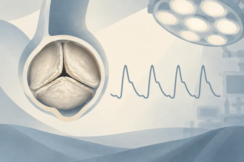
Suppose the examiner describes a 74-year-old with exertional dyspnoea and presyncope before elective total hip arthroplasty. Your first recommendation is to pause the operation. Establish whether the aortic stenosis (AS) is severe and whether it explains the symptoms, then seek Heart Team assessment. If symptomatic severe AS is confirmed and valve intervention is indicated and suitable, elective noncardiac surgery should follow valve treatment.
That is the decision at the centre of this ANZCA Final viva case. The trap is to jump straight to monitoring and vasopressors—or to declare that every patient with severe AS must have every operation cancelled. Symptoms, ventricular function, surgical urgency and the risks of valve treatment and noncardiac surgery all matter.
Confirm the lesion and make the symptoms count
Clarify when the symptoms began, whether they are worsening, and whether breathlessness or presyncope occurs with exertion. Ask about angina, syncope and heart failure. Establish functional capacity and whether reduced activity might be masking symptoms. Breathlessness can have other causes, but exertional presyncope in a patient with AS warrants careful assessment.
Review a recent transthoracic echocardiogram (TTE). Severe-range measures include a peak aortic jet velocity of at least 4.0 m/s, a mean gradient of at least 40 mmHg and an aortic valve area (AVA) of 1.0 cm² or less. These measures can disagree. Recheck the left ventricular outflow tract measurement and Doppler windows, then interpret the results alongside flow, left ventricular ejection fraction (LVEF) and the clinical picture. Do not select the single measurement that best fits your preferred plan.
Look beyond the valve area. Left ventricular function, hypertrophy and filling, right ventricular function, pulmonary pressures, coronary disease and other valve lesions all affect perioperative risk. Review the electrocardiogram, comorbidities, frailty and the proposed operation. The examiner wants an integrated assessment, not a decision based on one echocardiographic number.
Let urgency—not the diagnosis alone—set the pathway
For an elective operation that can wait, pause and discuss the case with the Heart Team, including cardiology, cardiac surgery, anaesthesia and the surgical team. The team can assess whether surgical or transcatheter aortic valve intervention is appropriate and how its timing affects the planned operation.
The recommendation changes when surgery cannot wait. Weigh the harm of delay against the risk of proceeding with the stenotic valve. For life-saving or time-sensitive surgery, careful haemodynamic monitoring, avoidance of rapid changes in volume status and prompt treatment of arrhythmias are central. Before urgent high-risk surgery in a patient with critical AS, transcatheter valve intervention or balloon aortic valvuloplasty may be considered when feasible. Balloon valvuloplasty can cause severe acute aortic regurgitation. Some urgent low- or intermediate-risk operations can proceed with close monitoring and a clear plan for instability.
Symptoms do not make every pathway identical. An asymptomatic patient with preserved ventricular function may be able to proceed with an elective low-risk operation after appropriate evaluation. Higher-risk surgery, ventricular dysfunction, symptoms or expected major haemodynamic changes alter that assessment. Explain what would make you proceed; do not rely on the absolute rule that “severe AS means no surgery.”
If surgery proceeds, protect perfusion
State the haemodynamic priorities before naming an anaesthetic technique: avoid hypotension, tachycardia and abrupt changes in circulating volume, and preserve sinus rhythm when possible. A hypertrophied left ventricle may tolerate sudden changes in filling or systemic pressure poorly. Fluid excess is not the answer to every low blood pressure; pulmonary congestion is also dangerous.
Match monitoring to the patient and the operation. For a symptomatic patient proceeding to major surgery, consider an arterial catheter before induction when rapid pressure changes or instability are likely; it is not an automatic requirement for every patient with AS. Ensure reliable intravenous access and have vasopressor support immediately available. Transoesophageal echocardiography may help in complex cases or unexplained instability. A pulmonary artery catheter is not routine.
No induction agent or technique removes the underlying risk. Titrate the anaesthetic to avoid abrupt vasodilation or myocardial depression, and anticipate the haemodynamic response to airway management. The choice of technique matters less than a clear plan to recognise and treat hypotension promptly.
Treat post-induction hypotension as a threat to perfusion
Imagine that pressure falls after induction, the heart rate rises and the electrocardiogram shows new ST depression. Treat this as possible myocardial ischaemia, not as an ordinary transient dip. Pause surgical stimulation, call for help and assess the patient systematically: confirm the arterial trace and rhythm, check oxygenation and ventilation, and look for recent anaesthetic dosing, vasodilation, reduced filling, bleeding or arrhythmia.
Restore pressure promptly with a titrated vasopressor chosen for the haemodynamic picture. If volume loss is likely, give fluid cautiously and reassess rather than loading a stiff ventricle blindly. Reduce excessive anaesthetic depth, address tachycardia or arrhythmia, and assess the response after each intervention.
If hypotension or ischaemic changes persist, escalate. Use echocardiography if available to assess ventricular filling and function, consider myocardial ischaemia or acute heart failure, and involve senior anaesthesia and cardiology support. The examiner is listening for a sequence: recognise the threat, correct the cause, restore perfusion and reassess.
Carry the plan into recovery
Haemodynamic risk continues after extubation. Arrange postoperative care at a level suited to the operation and the patient’s stability, with ongoing observation for hypotension, tachycardia, arrhythmia, heart failure and myocardial ischaemia. Maintain careful fluid balance and provide effective analgesia. If valve intervention has occurred, coordinate the postoperative plan with the treating team.
A concise viva answer gives the examiner a clear route through your reasoning. The guide to structuring an ANZCA Final viva answer offers a framework; the ANZCA Final Examination guide covers examination details. For further oral-case practice, the Final/Advanced Anesthesia Structured Oral Examination (SOE) provides structured viva practice.
The sentence to leave with the examiner is simple: confirm whether severe AS explains the symptoms, defer elective surgery for Heart Team assessment when appropriate, and, if surgery cannot wait, protect perfusion from induction through recovery.
Frequently asked questions
Should elective noncardiac surgery be delayed in symptomatic severe aortic stenosis?
Pause the elective operation, confirm the severity of stenosis and whether it explains the symptoms, and arrange Heart Team assessment. If valve intervention is indicated and suitable, it should precede elective surgery. The plan changes when surgery cannot wait.
What echocardiographic findings indicate severe aortic stenosis?
Severe-range measures include peak aortic jet velocity of at least 4.0 m/s, mean gradient of at least 40 mmHg and aortic valve area of 1.0 cm² or less. Interpret the measurements together with flow, ventricular function and study quality; investigate discordant results rather than relying on one number.
What is the best induction agent for severe aortic stenosis?
There is no single best agent. Use a controlled, titrated technique, with close blood pressure monitoring and vasopressor support available. Tailor the plan to ventricular function and haemodynamic reserve.
What are the main anaesthetic goals in severe aortic stenosis?
Maintain perfusion and stable blood pressure, avoid tachycardia and abrupt changes in circulating volume, and preserve sinus rhythm when possible. Avoid both dehydration and fluid overload.