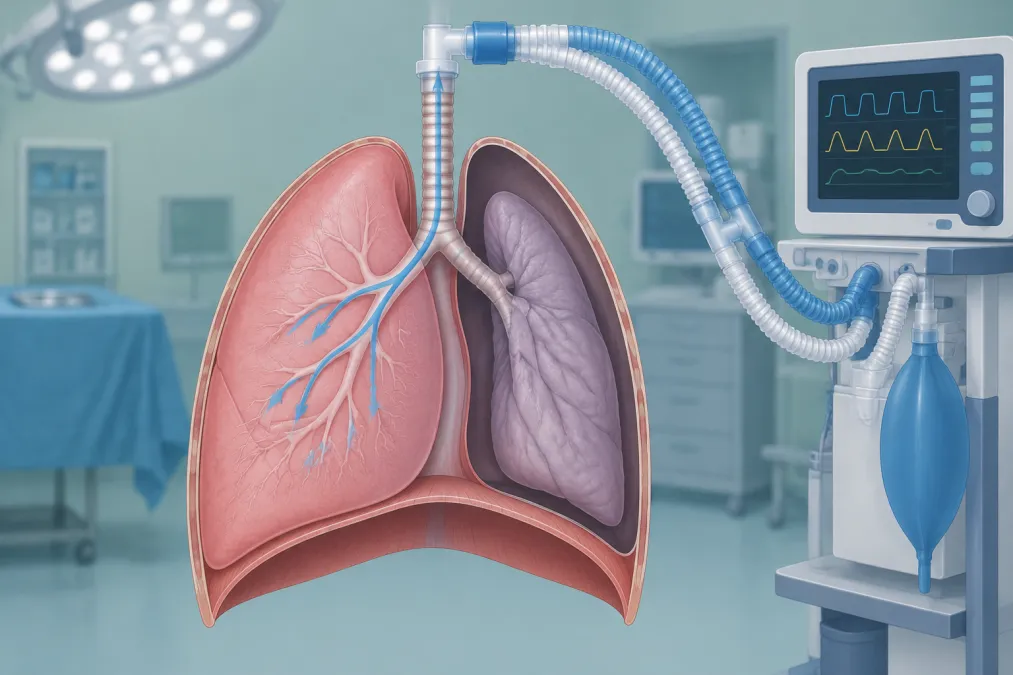
The saturation alarm sounds just as the surgeon asks for a quieter field. One lung is being ventilated; blood is still flowing through the other. That mismatch is the starting point for understanding one-lung ventilation (OLV)—and for responding when oxygenation falls.
Use a low tidal volume based on predicted body weight (PBW), individualize positive end-expiratory pressure (PEEP), and confirm the lung-isolation device with bronchoscopy. When hypoxaemia develops, a change in ventilation mode is not a rescue plan. First protect oxygenation; then identify a device problem, address the ventilated lung’s mechanics and haemodynamics, and consider how the surgical field limits shunt-reduction measures.
Why oxygenation falls during one-lung ventilation
Ventilation to one lung stops, but perfusion to the nonventilated lung continues. Blood passing through it returns to the left heart without being oxygenated, creating an obligatory right-to-left shunt. Hypoxic pulmonary vasoconstriction (HPV) narrows vessels supplying the nonventilated lung and redirects some blood flow toward the ventilated lung. It reduces the shunt; it does not eliminate it.
Gas exchange can worsen further if the ventilated lung develops atelectasis or low ventilation–perfusion regions. Anaesthesia, neuromuscular blockade, lateral positioning and opening the chest all affect lung volumes and mechanics. Secretions, bronchospasm, or displacement of a double-lumen tube (DLT) or bronchial blocker can add a correctable problem to the expected shunt.
Haemodynamics matter too. Low cardiac output can worsen arterial oxygen tension when shunt is present. Blood loss and anaemia can reduce oxygen delivery even when the saturation reading itself does not change. A falling saturation is therefore not automatically a ventilator-setting problem.
Set a protective baseline, then individualize
A common OLV tidal-volume starting range is 4–5 mL/kg PBW. PBW is calculated from height and sex, not actual body weight; using actual weight in a patient with obesity can lead to an unnecessarily large volume for the ventilated lung. Reassess the delivered volume as mechanics change.
A starting PEEP near 5 cm H₂O is reasonable for many patients, but it is a starting point, not a prescription for every lung. Titrate against oxygenation, compliance, driving pressure—the plateau pressure minus PEEP—and blood pressure. In obstructive lung disease, look for expiratory flow that has not returned to baseline and consider intrinsic PEEP before adding external PEEP. Excessive PEEP can overdistend the ventilated lung, impair venous return or worsen ventilation–perfusion matching.
Monitor tidal volume, plateau pressure, driving pressure, compliance, oxygenation and blood pressure together. If dependent-lung atelectasis is likely, a recruitment manoeuvre may help; reassess the patient’s response rather than applying it as a routine reflex.
The PROTHOR trial compared PEEP of 10 cm H₂O with periodic recruitment manoeuvres against PEEP of 5 cm H₂O without routine recruitment manoeuvres. The higher-PEEP strategy did not significantly reduce postoperative pulmonary complications. It was associated with more intraoperative hypotension and arrhythmias, while hypoxaemia rescue manoeuvres were more frequent with the lower-PEEP strategy. The result argues against treating fixed high PEEP as a universal solution; it does not mean PEEP should be avoided.
Volume control or pressure control?
In volume-controlled ventilation (VCV), the ventilator targets a set tidal volume, while airway pressure changes with compliance and resistance. In pressure-controlled ventilation (PCV), the inspiratory pressure is set, so delivered tidal volume can change as mechanics or surgical conditions change. With PCV, watch the exhaled tidal volume closely, particularly during lung or chest manipulation.
PCV may produce a lower displayed peak airway pressure. That reading alone does not establish lower alveolar stress or prove that oxygenation will improve. A small meta-analysis found a modest oxygenation difference in favour of PCV, but a later randomized trial found no difference in postoperative pulmonary complications among VCV, PCV and pressure control with volume guarantee. Choose the mode that reliably delivers the intended protective tidal volume and lets you follow pressure and mechanics. Do not switch modes expecting the shunt to disappear.
A falling saturation: work in order
For abrupt, severe or worsening hypoxaemia, tell the surgeon, ask for the operation to stop, increase inspired oxygen to 1.0 and promptly resume two-lung ventilation if possible. Do not delay urgent oxygenation while pursuing a detailed diagnosis. Once the situation is stable enough for a measured assessment, work through the likely causes:
- Check the circuit and airway setup. Confirm oxygen delivery, circuit connections, clamps, capnography and the saturation signal. Ask the surgeon to pause if that makes access safer.
- Confirm lung-isolation device position with bronchoscopy. Recheck after the lateral turn, surgical manipulation, a change in oxygenation or an unexpected rise in airway pressure. Look for displacement, lobar obstruction, cuff problems and secretions; suction as needed.
- Optimize the ventilated lung. Assess for bronchospasm, obstruction, atelectasis, gas trapping or pneumothorax. If dependent-lung collapse is likely, consider recruitment and carefully titrated PEEP. After each intervention, reassess tidal volume, pressures, oxygenation and blood pressure.
- Review haemodynamics and oxygen delivery. Look for blood loss, hypotension, low cardiac output or significant anaemia, and treat the cause when present. Avoid reflex fluid loading; excessive fluid can worsen lung water.
- Reduce shunt if hypoxaemia persists. If the surgical field permits, apply continuous positive airway pressure (CPAP) of 2–5 cm H₂O with an inspired oxygen fraction of 1.0 to the nondependent, nonventilated lung. Oxygen insufflation or intermittent two-lung ventilation are alternatives. Agree the manoeuvre with the surgeon because reinflation can compromise exposure. For severe or continuing desaturation, return to two-lung ventilation.
Work the reasoning through
Imagine saturation falls after the patient is turned lateral and peak airway pressure rises. A mode change may alter the displayed pressure, but it does not answer the key question: is the dependent lung being ventilated, and is the isolation device still correctly positioned? Increase inspired oxygen, alert the surgeon and check the circuit. Then confirm the DLT or blocker with bronchoscopy and suction if required.
If the device is correctly positioned but the dependent lung is poorly compliant, consider atelectasis, bronchospasm or gas trapping. Treat the likely cause and reassess after any recruitment manoeuvre or PEEP adjustment. If device position and lung mechanics are satisfactory but oxygenation remains poor, consider shunt and the surgical options: CPAP to the nonventilated lung or temporary two-lung ventilation may help more than another mode switch.
The viva traps worth avoiding
Separate peak pressure from lung stress, and device malposition from expected shunt. PCV may lower peak pressure, but it does not guarantee a safer delivered tidal volume or better oxygenation. Calculate tidal volume from PBW, not actual body weight. Individualize PEEP and reassess its effect on lung mechanics and blood pressure.
For oral exam practice, the Final/Advanced Anesthesia Structured Oral Examination bank offers a place to rehearse a clear, spoken rescue sequence.
In OLV, the order matters: protect oxygenation, check the device, address the ventilated lung and oxygen delivery, then reduce shunt or restore two-lung ventilation. A mode switch may change the pressure trace. It does not, by itself, fix hypoxaemia.
Frequently asked questions
What tidal volume should you use during one-lung ventilation?
A common starting range is 4–5 mL/kg predicted body weight (PBW). Adjust ventilation to the patient’s lung mechanics and gas exchange; do not calculate the volume from actual body weight.
Is pressure-controlled ventilation better than volume-controlled ventilation during one-lung ventilation?
Neither mode is a reliable hypoxaemia rescue by itself. Pressure control may lower displayed peak airway pressure, but you must monitor delivered tidal volume and the patient’s response. A mode change does not correct device malposition or shunt.
What PEEP should you use during one-lung ventilation?
A starting PEEP near 5 cm H₂O is reasonable for many patients, but it should be individualized against oxygenation, lung mechanics, blood pressure and any intrinsic PEEP.
What should you do first when oxygen saturation falls during one-lung ventilation?
For abrupt, severe or worsening hypoxaemia, alert the surgeon, increase inspired oxygen and promptly resume two-lung ventilation if possible. Once the patient is stabilized, check the circuit and confirm lung-isolation device position with bronchoscopy.
References
Show 5 references
- Hypoxaemia during one lung ventilation
- Effects of intraoperative higher versus lower positive end-expiratory pressure during one-lung ventilation for thoracic surgery on postoperative pulmonary complications (PROTHOR): a multicentre, international, randomised, controlled, phase 3 trial
- Comparison of pressure-controlled ventilation with volume-controlled ventilation during one-lung ventilation: a systematic review and meta-analysis
- Effect of ventilation mode on postoperative pulmonary complications following lung resection surgery: a randomised controlled trial
- Lung-protective ventilation for the surgical patient: international expert panel-based consensus recommendations